University College London’s Human Organ Atlas lets anyone explore human organs in stunning 3D, from full organs down to individual cells, through a free, browser-based platform designed for researchers, educators, and curious minds
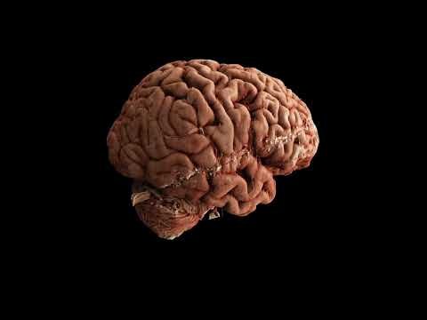
University College London (UCL) has launched the Human Organ Atlas, a revolutionary 3D platform that lets users explore organs such as the brain, heart, lungs, and kidneys, all from a standard web browser. Using cutting-edge imaging technology, the platform allows zooming from entire organs down to single cells, providing an unprecedented resource for medical research, education, and public discovery. Dubbed the “Google Earth for the human body,” this free, interactive tool opens up the human body like never before.
The paper is published in Science Advances.
UCL’s Human Organ Atlas reveals whole organs in 3D down to near-cellular detail
The Human Organ Atlas is powered by an advanced X-ray imaging method called Hierarchical Phase-Contrast Tomography (HiP-CT), developed at the European Synchrotron (ESRF) in Grenoble, France. The method uses the ESRF’s Extremely Brilliant Source – a new generation of synchrotron source, which is up to 100 billion times brighter than conventional hospital CT scanners.
This tool allows researchers to scan entire intact donated organs non-destructively, then zoom in to near-cellular resolution.
The Atlas was developed during the COVID-19 pandemic and played a key role in discovering the unseen microscopic vascular injury in the lungs of patients who died from COVID-19. Since its creation, the researchers wanted the Human Organ Atlas to be an open, shared scientific infrastructure at a global scale, operating as a resource for researchers, doctors, educators and anyone interested in the human body.
Professor Peter Lee (UCL Department of Mechanical Engineering), principal investigator of the Human Organ Atlas beamtime, said: “To create the Human Organ Atlas, we brought together scientists and medics from nine institutes worldwide. This grouping is continuing to expand, helping gain new insights into diseases from osteoarthritis to heart disease and changing how we learn about the human body.”
UCL’s Human Organ Atlas gives scientists and students 3D access to dozens of organs
The Human Organ Atlas currently provides access to:
- 62 organs, 319 full 3D datasets from 29 donors
- 12 organ types, including brain, heart, lung, kidney, liver, colon, eye, spleen, placenta, uterus, prostate and testis
- Multiscale scans, from whole-organ views down to near-cellular resolution (routinely down to 2 µm, as fine as 0.65 microns for some organs)
To make the data usable worldwide, the portal provides:
- Interactive browser-based visualisation (no special software required)
- Downloadable datasets at multiple resolutions
- Tutorials and software tools for analysis
- Regular addition of new data
Dr Alexandre Bellier, based at Grenoble Alpes University Hospital, said: “Students can explore organs in 3D, scroll through anatomical sections, and zoom into internal tissue detail. It creates an immersive, exploratory alternative to classic anatomy diagrams, helping learners build a clearer spatial understanding of complex structures. For both teachers and students, it fundamentally shifts anatomy learning from static description to guided, interactive discovery.”
Looking to the future, the team plans to expand the collection over the next few years, adding more organs, more samples, and new tools, while growing an open community around the data.
Dr Tafforeau said: “We are opening a new window into the inner architecture of the human body. After 6 years of efforts, we are still only at the beginning. Currently, we work on isolated organs, but in the future, we expect to develop the technique to be able to image complete human bodies with a resolution 10 to 20 times higher than what is possible today. Such data could transform how anatomy is studied and understood.”